
Researcher developing noninvasive imaging for anal cancer screening
The new technology will make use of automated image analysis algorithms to support future AI tools and faster delivery of results.
A University of Arizona Comprehensive Cancer Center researcher has received a $1.4 million grant from the National Institutes of Health to develop a novel imaging device that improves early detection and diagnosis of anal precancer.
Dongkyun Kang, PhD, an associate professor of optical sciences and biomedical engineering at the U of A Wyant College of Optical Sciences, hopes the technology he is developing will help address rising anal cancer rates. Mortality due to anal cancer increased by nearly 6% per year between 2014 and 2020, according to Kang.
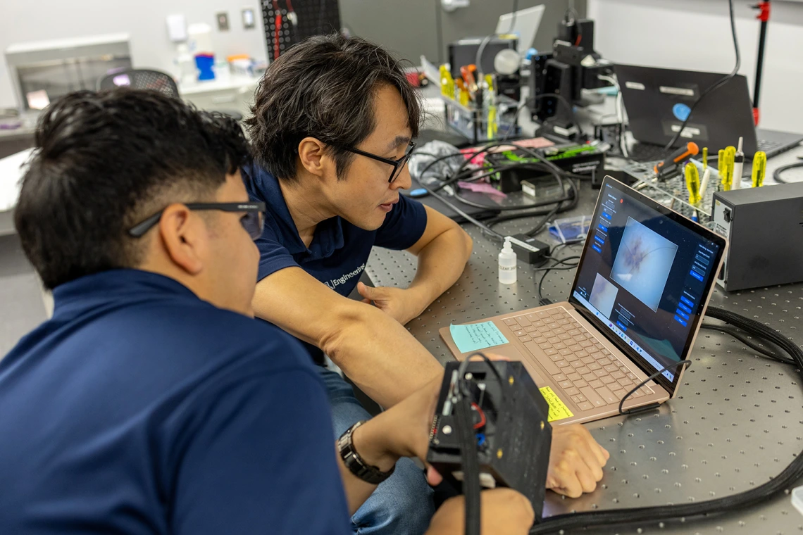
His lab will develop a probe-based light sheet microscope that allows clinicians to examine anal tissue in real-time, with high-resolution, noninvasive imaging.
“Our goal is to enhance early detection of anal cancer by giving clinicians a powerful new tool for examining suspicious lesions during screening,” said Kang. “The device could increase diagnostic accuracy, reduce unnecessary biopsies and ultimately improve outcomes for patients at high risk.”
This tool will address a critical gap in early screening methods. Until recently, there were no standard screening guidelines for anal cancer, largely due to limitations in diagnostic methods. Traditional, high-resolution anoscopy, currently used to guide biopsies, has variable accuracy and requires highly trained specialists. A 2022 anal cancer outcomes research study showed that early treatment of precancerous anal lesions, known as high-grade squamous intraepithelial lesions, can significantly reduce cancer risk, creating an urgent need for improved diagnostic tools.
Kang’s lab previously demonstrated the potential of scattering-based light sheet microscopy for identifying anal lesions in tissue samples. In this new four-year project, Kang and his team will miniaturize and refine the technology into a probe-based format suitable for use during live patient exams.
How it works
The scanning device is roughly the diameter of standard anal biopsy forceps and provides detailed images of tissue structures with cellular-level resolution. Clinicians can use these images to better target biopsies, potentially avoiding unnecessary removal of benign or low-grade lesions. The system also offers unique advantages for clinical training and real-time lesion monitoring.
“The device will allow for image-guided biopsy, reducing patient discomfort and increasing diagnostic confidence,” Kang said. “It can also provide immediate visual feedback for clinicians in training and help monitor suspicious areas over time. This is especially important in high-risk populations who may face barriers to follow-up care.”
The project includes three aims:
Develop and validate the scanning device for in vivo clinical use, ensuring it meets performance benchmarks for resolution, size, and usability
Conduct a clinical study involving 70 patients to compare the new device’s image findings with traditional pathology, measuring the accuracy, sensitivity, and specificity of the device for diagnosing various types of tissue
Build automated image analysis algorithms to support rapid and consistent interpretation of images by clinicians and future AI tools
“This grant enables us to bridge a significant technological and clinical gap in cancer diagnostics,” said Kang. “Our work could have broad impact not only on anal cancer screening, but also on other cancers like cervical and oral cancers that require evaluation of epithelial cell structures.”
A platform for broader cancer prevention
Kang envisions the new platform extending beyond anal cancer, offering a compact, cost-effective solution for point-of-care screening in a variety of cancers and healthcare settings. The team hopes this technology will eventually support same-day treatment decisions and expand access to high-quality diagnostics in a wide range of healthcare environments.
“The work Dr. Kang is leading represents a powerful blend of convergent science. This matches engineering innovation and the clinical needs of patients with the latest emergent data,” said Dan Theodorescu, MD, PhD, director of the University of Arizona Comprehensive Cancer Center and the Nancy C. and Craig M. Berge Endowed Chair. “It’s an outstanding example of how convergent science can reshape cancer detection and care. We look forward to translating these findings and taking them to the clinic with our partners at Banner Health.”