
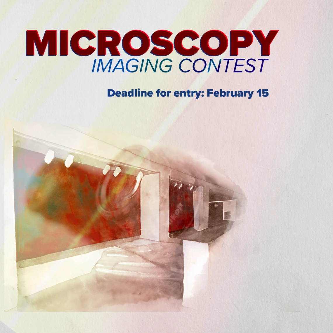
CONTEST RULES
Deadline for submission: February 15
Images can have any basic research or clinical focus
Up to 3 submissions per person
IMAGE GUIDELINES
Submitted as a .ND2 raw or .Tiff file
Acquired on a MSR (Nikon) microscope Still images only (no timelapses)
CONTEST PRIZES
Top 3 rated submissions will:
Be printed to be displayed in the UA Cancer Center Atrium
Receive a personal, mounted print of the submitted image
Receive *cash and Nikon-themed prizes
Submissions will be judged by a panel of researchers campus-wide
*Cash prizes minus applicable taxes will be delivered for UA employees via paycheck. Non UA employees will receive a check less applicable taxes.
How to submit and name your contest entries:
Each user will create a folder on their computer containing the images. A folder should be created even if there is only a single image.
Name each image file with the following information:
User Name_Sample Type (i.e. tissue type, cell line or cell type name, organoid)_Image Number (if needed)
Example:
- Marco Padilla_Mouse Brain Tissue_1
- Marco Padilla_Mouse Brain Tissue_2
- Marco Padilla_Tumor Microarray
Include one short text document that has the following details for each image:
Example:
- Marco Padilla_Mouse Brain Tissue_1 and _2
- This image is of fixed mouse brain tissue with the following stains: Nuclei = DAPI, Neurons = AF488, Amyloid Beta = AF546, Microglia = AF647
- Marco Padilla_Tumor Microarray
- This image is a polarized brightfield image of a breast cancer tumor microarray sample.
Beyond describing what is being shown in each channel, no experimental details are required.
Upload all entries to the following link:
https://arizona.app.box.com/f/1661a16afcc9407bbc08c326d7768263
Questions? Contact: Marco Padilla-Rodriguez @ Marco@arizona.edu