Flow Cytometry and Immune Monitoring Shared Resource
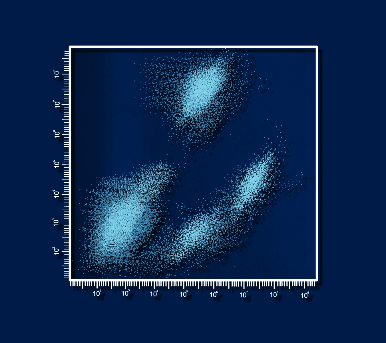
Flow cytometry is a powerful tool that measures the functional and structural characteristics of heterogeneous mixtures of cells and particles in suspension based on their ability to scatter light. The cell sorting function separates these cells physically into their different classes. Researchers have the capability to analyze and sort cells by differences in physiology, metabolism, morphology and other characteristics. The ability to distinguish different cell types is limited only by the ability to attach specific fluorescent markers to the cells.
Flow Cytometry
Our state-of-the-art flow cytometry instrumentation is available for data acquisition, analysis, and cell sorting, and the technical expertise to interpret results, and develop methods. We offer information about new techniques, and applications of flow cytometry through workshops and seminars, and provide training to interested facility users who wish to run their own samples. Individual consultation services are available to discuss the specifics of each project.
Immune Monitoring
The Immune Monitoring Shared Resource provides comprehensive immune profiling, sample preparation, and clinical trial support. Our mission is to provide technical expertise to clinical investigators and research scientists who want to characterize the immune system by offering up-to-date immune monitoring services for clinical and translational studies.
About the FCIMSR:
We offer flow cytometry and cell sorting instrumentation and expertise, as well as immune profiling of clinical samples. These combined cores support academic research all the way from basic science to translation and clinical trials.um/plasma extraction, and a full spectrum of immunological services. We also offer custom assays upon request and will work with interested investigators to help get their projects up and running. Biobanking services are also available upon request.
We offer training, assisted, unassisted, and technician-operated modalities for all our instruments and will meet investigators at their desired level of engagement. All services are available during regular business hours (M-F 8am - 4pm) with 24-hour access to select instruments.
Our Services:

Flow cytometry is a powerful tool that measures the functional and structural characteristics of heterogeneous mixtures of cells and particles in suspension based on their ability to scatter light. Researchers have the capability to analyze and sort cells by differences in physiology, metabolism, morphology and other characteristics.

Our services include multispectral flow cytometry, metabolic analysis, blood services such as PBMC isolation and serum/plasma extraction, and a full spectrum of immunological services.
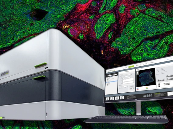
The FCIMSR houses a NanoString GeoMx Digital Spatial Profiler (DSP) which is a spatial biology instrument that enables morphology-driven, high-plex profiling on a single FFPE or fresh frozen tissue section. DSP technology combines standard immunofluorescence techniques with nCounter optical barcoding quantification. This technology allows for spatially resolved, high-plex (10s -10,000s) digital quantitation of proteins or mRNA while maintaining integral information about tissue architecture and spatial relationships between cellular networks.
Equipment
- The FACS Canto II (BD Biosciences, San Jose, CA)
- is an analyzer which can acquire data from eight fluorescence detectors using three lasers: 405nm, 488nm, and 640nm.
- The FACS Aria III (BD Biosciences, San Jose, CA)
- is a cell sorter equipped with 5 lasers: 488nm, 633nm, 561nm, 405nm and 355nm, and offers 17 fluorescence detectors. The sorting function of this instrument enables the researcher to physically separate the individual cells and particles from a mixed population. Viable cells can be recovered for further study or culture. The addition of a Baker BioProtect III biosafety cabinet, in which the Aria is housed, allows for sorting of certain live human tissue and BSL2 agents.
- The LSRII (BD Biosciences, San Jose, CA)
- is an analyzer capable of acquiring and analyzing data from 16 fluorescence parameters and has five lasers: 355nm, 405nm, 488nm, 532 nm and 640nm.
- The Cytek Aurora (Cytek Biosciences, Fremont, CA)
- is a multispectral flow cytometer. This instrument is capable of 64 channels of detection across 5 lasers using up to 40 colors in a single multicolor sample. Laser wavelengths include 355nm, 405nm, 488nm, 561nm, and 640nm. This cytometer also comes equipped with a 96 well plate loader for high throughput cytometry applications.
- The ImagestreamX MarkII
- is an imaging cytometer, which uses flow cytometry technology to acquire microscopic images of fluorescence data from thousands of individual cells in liquid suspension. It has 10 fluorescence channels, using four lasers: 405nm, 488nm, 532nm, and 640nm.
- The Fortessa (BD Biosciences, San Jose, CA)
- which has been in the care of the Nikolich-Zugich laboratory for many years is now administered by the FCIMSR. It can be found in the Medical Research Building, room 240K2, directly across from the LSRII flow cytometer. The Fortessa is functionally identical to the LSRII, but newer, with 7 detectors off the violet laser compared to only 4 detectors on the LSR.
- The Attune (ThermoFisher)
- is a 14 ‘color’ flow cytometer with an exceptionally fast tube and plate sample rate. It uses sound waves to focus the sample stream into the laser intercepts, so its data precision is almost unaffected by flow rate, unlike a conventional flow cytometer which uses fluid dynamics to focus the sample which cannot maintain precision at even moderate flow rates.
- The Seahorse XFe96 Metabolic Analyzer (Agilent Technologies, Santa Clara, CA)
- is used to measure the oxygen consumption rate (OCR) and extracellular acidification rate (ECAR) of live cells in a 96-well plate format. OCR and ECAR rates are key indicators of mitochondrial respiration and glycolysis as well as ATP production rate. Together these measurements provide a systems-level view of cellular metabolic function in cultured cells and ex vivo samples
- The QuantStudio 3D Digital PCR System (Applied Biosystems, Foster City, CA)
- is a simple, affordable platform that uses sealed-chip technology, providing a streamlined, reliable, and robust method for performing digital PCR. The turn-key chip-based workflow allows for loading the chip, sealing and proceeding to amplification. Featuring 20,000 nanoscale reaction wells per chip, this platform enables more controlled sample loading and less reaction dropout than droplet-based approaches, ensuring higher precision for absolute quantification
- The BioTek Cytation 1 Cell Imaging Multi-Mode Reader (BioTek Instruments Winooski, VT)
- combines high sensitivity filter-based fluorescence and high contrast brightfield imaging with conventional multimode detection. The Cytation 1 can interface with the Seahorse instrument to allow for real-time normalization for Seahorse assays.
- The NanoString GeoMx Digital Spatial Profiler (DSP) (Nanostring Seattle, WA)
- is a spatial biology instrument that enables morphology-driven, high-plex profiling on a single FFPE or fresh frozen tissue section. DSP technology combines standard immunofluorescence techniques with nCounter optical barcoding quantification. This technology allows for spatially resolved, high-plex (10s -10,000s) digital quantitation of proteins or mRNA while maintaining integral information about tissue architecture and spatial relationships between cellular networks
- The Nanodrop One (ThermoScientific)
- is a compact, stand-alone UV-Visible spectrophotometer developed for micro-volume analysis of purified nucleic acids and a wide variety of proteins. The patented sample retention system enables the measurement of highly concentrated samples without the need for dilution. The Nanodrop One comes with preloaded software and a touchscreen display.
Flow Cytometry Standard Operating Procedures HIMF Standard Operating Procedures
To build a project, get a quote, or order a service within iLab:
- Step 1: Select “About Our Core” to learn more about this Shared Resource.
- Step 2: Login or sign up for an iLab account at the University of Arizona. For login help, email iLab-support@agilent.com.
- Step 3: Once the login step is completed, obtaining prices, services and scheduling is available within the iLab system.
Please remember to acknowledge the Cancer Center Support Grant (P30 CA023074) when publishing manuscripts or abstracts that utilized the services of the University of Arizona Cancer Center’s Shared Resources and/or were derived from CCSG pilot funds. Suggested language: "Research reported in this [publication/press release] was supported by the National Cancer Institute of the National Institutes of Health under award number P30 CA023074."